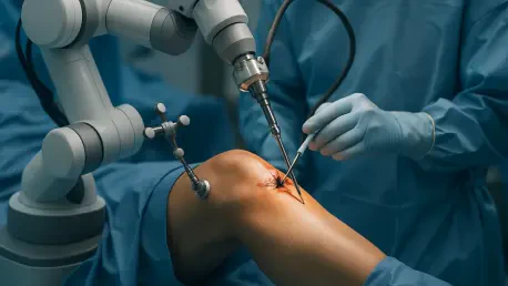
Imagine a world where knee surgeries are not only faster but also far more accurate, reducing recovery times and improving long-term outcomes for patients worldwide. This vision is becoming a reality thanks to a groundbreaking advancement in medical technology driven by artificial intelligence (AI). Specifically, a new deep learning model developed by esteemed researchers at Seoul National University is transforming how orthopedic surgeons approach knee surgery. By focusing on the precise measurement of the tibial posterior tilt angle—a critical factor in joint stability and the success of artificial joint implants—this AI innovation promises to standardize a process that has long been plagued by inconsistency. As the medical field grapples with the challenges of varying X-ray imaging practices across institutions, this technology offers a unified solution that could redefine surgical planning and patient care on a global scale.
Transforming Orthopedic Diagnostics with AI Precision
The challenge of measuring the tibial posterior tilt angle has historically hindered orthopedic surgery due to the lack of a standardized method. Variations in X-ray imaging techniques across different medical facilities often result in inconsistent measurements, which can compromise the reliability of clinical research and surgical outcomes. This inconsistency poses a significant barrier to ensuring knee joint stability and the longevity of artificial joints after surgery. Enter the AI model developed by Professors Noh Doo-hyun and Kim Sung-eun, which automates the measurement process with unprecedented accuracy. By recognizing six key anatomical landmarks on knee X-ray images, the model calculates the joint line and central axis of the tibia to determine the posterior slope. This automated approach not only eliminates human error but also sets a new benchmark for precision in orthopedic diagnostics, paving the way for more reliable surgical planning.
Beyond accuracy, the efficiency of this AI model stands out as a game-changer for medical professionals. Traditional manual measurements of the tibial posterior tilt angle often take specialists an average of 26.1 seconds per image, a process that can become time-consuming when handling large volumes of cases. In stark contrast, the AI completes the same task in just 2.5 seconds—over ten times faster. This remarkable speed is backed by data from over 10,000 knee X-ray images, ensuring the model’s robustness. Furthermore, it achieves a perfect 100% intra-observer correlation coefficient, meaning it delivers consistent results every time, unlike manual methods which peak at 95%. With an inter-observer correlation coefficient of 91%, the AI’s measurements align closely with those of experienced specialists, proving its reliability as a diagnostic tool and highlighting its potential to streamline workflows in busy clinical environments.
Setting a Global Standard for Knee Surgery Measurements
One of the most promising aspects of this AI technology is its potential to establish a universal standard for measuring the tibial posterior tilt angle across diverse populations. A follow-up study involving 289 Norwegian patients demonstrated an impressive 80% correlation with specialists’ measurements, underscoring the model’s adaptability beyond the population it was initially developed for. This cross-ethnic applicability suggests that the AI could be implemented in hospitals worldwide, addressing the long-standing issue of variability in medical imaging practices. As knee surgeries become increasingly common due to aging populations and active lifestyles, having a consistent measurement tool could significantly enhance patient outcomes by ensuring that surgical interventions are based on accurate and comparable data, regardless of geographic location.
The implications of this innovation extend far beyond individual patient care, influencing the broader landscape of orthopedic research and clinical practice. Research Professor Kim Sung-eun has highlighted the successful validation of this domestically developed medical AI across multiple ethnic groups, expressing optimism about its future versatility. Plans are underway to conduct additional studies to further refine and expand the model’s applications. Published findings in the Orthopaedic Journal of Sports Medicine reinforce the consensus that AI-driven tools like this one could revolutionize standardized medical measurements. By reducing variability and enhancing prognosis accuracy, the technology not only improves surgical precision but also contributes to the global advancement of orthopedic science, potentially shaping protocols and guidelines for years to come.
Pioneering the Future of Surgical Innovation
Reflecting on the strides made in knee surgery, the introduction of this AI model marked a turning point in how orthopedic challenges are addressed. Its ability to automate a previously inconsistent and labor-intensive process redefined efficiency, delivering results that are both rapid and reliable. Surgeons and researchers alike recognize the profound impact of achieving a 100% consistency rate, a feat that manual methods have never accomplished. This technological leap bridges gaps in clinical practice, ensuring that patients benefit from surgeries planned with unparalleled precision.
Looking ahead, the focus should shift to integrating this AI technology into everyday clinical settings worldwide. Hospitals and medical institutions must prioritize training staff to utilize such tools effectively, while ongoing research should aim to adapt the model for other complex measurements in orthopedic care. Collaboration between technologists and healthcare providers will be key to unlocking the full potential of AI, ensuring that innovations continue to evolve in response to real-world needs. By embracing these advancements, the medical community can build a future where surgical outcomes are consistently optimized for every patient.


