
Manual sample preparation, dilution, and reagent management create significant labor costs and potential for human error in contemporary laboratory environments. These foundational tasks underpin nearly every breakthrough in the life sciences, yet they often represent the most vulnerable links in the experimental chain. For researchers working at the cutting edge of oncology or regenerative medicine, the precision of a cell count is not merely a technicality but a prerequisite for the safety and efficacy of potential treatments. As laboratories transition into more automated frameworks, the reliance on subjective measurements is being replaced by data-driven insights. The precision required for modern bioprocessing necessitates a level of accuracy that transcends the capabilities of traditional microscopy. This evolution ensures that seeding densities are optimized and culture health is monitored with unprecedented clarity, providing a stable foundation for complex downstream applications that demand rigorous standardization across global research facilities.
The Legacy Bottleneck: Limitations of Manual Counting
For decades, the manual hemocytometer stood as the gold standard for cell counting, yet its reliance on human visual judgment introduced an unavoidable layer of subjectivity. Different operators, or even the same operator at different times of the day, might perceive cell boundaries and morphology with varying degrees of accuracy, leading to significant inter-lab variability. In an era where 2026-era research requires extreme precision, this lack of standardization has become a major hurdle for teams attempting to replicate findings across different geographical locations. The cognitive load required for manual counting also leads to fatigue, which further increases the likelihood of clerical errors and misinterpretations. Furthermore, the extensive hands-on time required for these procedures prevents skilled scientists from focusing on higher-value activities, such as experimental design and advanced data analysis, thereby slowing down the overall pace of therapeutic development.
Building on these procedural challenges, the use of chemical dyes like Trypan Blue presents a chemical limitation that modern labs can no longer afford to ignore. While these stains provide a binary live-or-dead assessment, they are inherently toxic to the cell population, necessitating that the sampled portion be permanently discarded. This loss is particularly damaging when working with rare primary cells or expensive engineered populations where every single cell is vital for downstream therapeutic use. Traditional staining methods also provide a relatively shallow view of cellular health, as they only identify membrane permeability after significant damage has already occurred. This reactive approach to viability assessment means that early-stage cellular stress often goes undetected, potentially leading to the use of compromised cells in sensitive experiments. The industry’s move toward stain-free alternatives is therefore driven by the dual need to preserve sample integrity and to gain more nuanced insights.
Technological Convergence: Digital Holographic Microscopy and AI
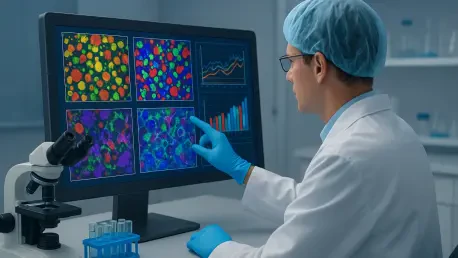
The transition to stain-free technology marks a revolutionary change in how scientists visualize and analyze living cells through Digital Holographic Microscopy (DHM). This technology functions by capturing quantitative phase information, recording how individual cells interact with and delay light waves. Unlike traditional brightfield microscopy that requires pigments or specialized filters to create contrast, DHM generates high-resolution digital holograms of cells in their native, undisturbed state. This approach completely eliminates the need for destructive chemical markers, allowing the entire cell population to remain viable and available for further experimentation or immediate therapeutic use. By removing the harsh environmental shifts associated with traditional staining, DHM provides a much more accurate representation of how cells behave in vivo, ensuring that the data collected is reflective of the culture’s true physiological condition during the analysis.
When this advanced imaging is integrated with machine learning, it transforms into a sophisticated analytical powerhouse capable of identifying cells with superior precision. Modern algorithms are trained to recognize the unique optical and morphological signatures of various cell types, allowing the system to distinguish between living cells, dead cells, and non-cellular debris automatically. This synergy enables the instantaneous calculation of cell concentrations and the assessment of viability without any manual intervention or human bias. By relying on physical properties such as refractive index and internal density rather than dye permeability, these systems provide a non-destructive alternative that preserves the integrity of even the most sensitive cell populations. The marriage of physical optics and computational intelligence represents a significant milestone in laboratory instrumentation, offering a level of depth that was previously impossible.
Advancing Diagnostics: From Binary Viability to Comprehensive Health
One of the most significant advantages of this new technology is the ability to view cell health as a continuous spectrum rather than a simple live-or-dead status. Traditional assays only provide a snapshot of whether a cell membrane is intact, often missing the critical early stages of cellular stress that precede death. In contrast, DHM-based systems like the CytoDirect can detect subtle shifts in size, shape, and internal density that indicate a change in metabolic state. This level of detail allows researchers to identify early signs of apoptosis long before a cell would take up a traditional stain, providing a much more nuanced understanding of culture vitality and long-term stability. This diagnostic depth is essential for researchers who must ensure that their cell populations are not just alive, but are functioning at their peak biological capacity for use in complex assays.
In the high-stakes world of biomanufacturing, understanding the potency of a cell population is just as vital as confirming its survival. By monitoring morphological changes in real time, scientists can make proactive decisions to adjust culture conditions or nutrient feeds before a population experiences a significant decline. This shift from reactive to proactive management is essential for optimizing the production of biologics and high-purity vaccines. The ability to see deeper into cellular behavior ensures that the cells used in critical treatments are of the highest possible quality and functionality. Furthermore, the objective nature of these measurements allows for the development of more accurate predictive models, which can be used to forecast the success of a batch based on early morphological trends. This predictive capability reduces the risk of batch failure and ensures a more consistent supply of vital medications.
Operational Excellence: Economic Drivers of Automation
The adoption of automated, stain-free counters brings immediate and practical benefits to laboratory management and overall workflow. By removing the time-consuming steps of preparation and incubation associated with staining, these systems generate results in a matter of seconds rather than minutes. This rapid speed allows for real-time monitoring of experiments, significantly accelerating the pace of discovery and allowing labs to process a much higher volume of samples each day. Moreover, the elimination of recurring costs for specialized consumables and toxic reagents improves the long-term budgetary health of the facility. The reduction in hazardous waste disposal costs also aligns with the broader industry trend toward sustainable and environmentally friendly laboratory practices. By streamlining the most repetitive parts of the workflow, facilities can allocate their resources more effectively toward innovation.
Automation also plays a crucial role in standardizing data across the scientific community by removing the human factor from the counting process. Whether a count is performed by a senior researcher or a junior technician, the results remain consistent and objective, which is critical for maintaining the integrity of large-scale clinical trials. This standardization is paired with the benefit of sample preservation; because the process is non-destructive, every cell can be returned to the culture vessel or used for downstream analysis. This is particularly vital in clinical settings where the starting material, such as patient-derived stem cells, is often limited and every single cell counts toward a successful outcome. The ability to conduct frequent, non-destructive checks allows for tighter control over the entire production process, ensuring that the final product meets the most stringent quality standards.
Navigating the Transition: Strategies for Long-Term Implementation
As the life sciences sector continues to evolve toward personalized medicine and advanced biologics, the limitations of legacy analytical methods have become increasingly apparent to strategic planners. The movement toward stain-free, automated analysis is a direct response to the need for greater analytical rigor and data fidelity in a competitive global market. Systems designed for this purpose act as force multipliers, allowing scientists to pivot their focus from repetitive manual tasks to high-level experimental design and sophisticated data interpretation. This evolution ensures that the tools used in the lab are just as advanced as the therapies they help create, bridging the gap between basic research and commercial application. By integrating these systems into existing laboratory information management systems, organizations can create a seamless flow of data that enhances transparency and traceability.
Organizations that successfully integrated these stain-free protocols recognized the immediate stabilization of their data pipelines and the subsequent reduction in experimental rework. To maximize the benefits of this technology, laboratories conducted thorough audits of their current manual bottlenecks and replaced them with validated automated workflows that prioritized non-destructive analysis. The transition required an initial investment in instrumentation, but the long-term gains in operational efficiency and sample recovery provided a rapid return on investment. Future-ready facilities also prioritized training programs that familiarized staff with the interpretation of morphological data, moving beyond simple concentration metrics toward a deeper understanding of cellular health. These proactive steps ensured that the data driving innovation remained both accurate and reproducible, securing a competitive advantage in the rapidly advancing landscape of biotechnology.